
Fig. 14.
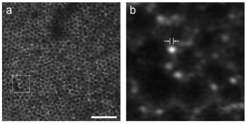
Retinal pigment epithelium and individual lipofuscin granules revealed in FAOSLO. (a) Individual RPE cells imaged using FAOSLO in macaque. Scale bar is 100 microns. (b) Outlined region from a showing individual lipofuscin granules; distance between arrowheads is 2 microns, on the order of the size expected for RPE granules.
From Rossi 2011.