
Fig. 15.
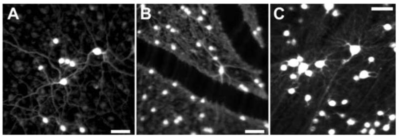
Fluorescence AOSLO images of primate retinal ganglion cells in vivo A), B) and C) Fluorescence AOSLO imaging revealed the morphology of retinal ganglion cells labeled with fluorophore (rhodamine dextran) in living monkey eye. The transverse resolution of the images is fine enough to resolve the individual dendrites. The fluorophore was introduced into the ganglion cells through retrograde labeling via injections in the lateral geniculate nucleus (LGN). Scale bar of 50 μm in all panels. [Panels A and C, Reproduced from Gray DC, Wolfe R, Gee BP, et al. (2008) In vivo imaging of the fine structure of rhodamine-labeled macaque retinal ganglion cells. Invest Ophthalmol Vis Sci 49:467-473, their Figures 1 and 5, with permission from Association for Research in Vision and Ophthalmology (Copyright 2008).]