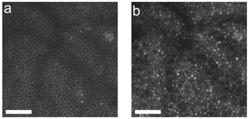
Fig. 19.

A. two-photon image of the cone mosaic in the living primate retina at 2.5 deg superior retina using 730 nm illumination from a Ti-Sapph laser with a pulse width of less than 70 fs. B. Reflectance image of the cone mosaic at the same location using 790 nm light. The bright spots in both images correspond, confirming the cone mosaic as the origin of the two photon signal. Scale bar, ?0 μm.
