Abstract
Background
Step-up exercise is one of the most commonly utilized exercises during rehabilitation of patients after both ACL injury and reconstruction. Currently, insurance providers increasingly required a trial of intensified rehabilitation before surgical reconstruction is attempted. The purpose of this study was to investigate whether this “safe” rehabilitation exercise in the setting of ACL deficiency can cause altered knee kinematics.
Methods
Thirty patients with unilateral ACL rupture were recruited for this study. The mean time from injury was 3.3 months. Tibiofemoral kinematics were determined during a step-up exercise using a combination of MRI, dual fluoroscopy and advanced computer modeling.
Findings
The ACL-injured knee displayed an average 5° greater external tibial rotation than the uninjured knee (p<0.05), during the last 30% of step-up. The ACL-injured knee also demonstrated on average 2.5 mm greater anterior tibial shift during the last 40% of stance phase (p<0.01). In addition, during the last 30% of stance the tibia of the ACL-deficient knee tended to shift more medially (~1 mm) as the knee approached full extension (p<0.01).
Interpertation
The data confirmed the initial hypothesis as it was found that ACL deficient knees demonstrated significantly increased anterior tibial translation, medial tibial translation and external tibial rotation towards the end of the step-up as the knee approached full extension. Intensive rehabilitation utilizing the step-up exercise in the setting of ACL deficiency can potentially introduce repetitive microtrauma by way of altered kinematics.
INTRODUCTION
Step-up exercise is one of the most commonly utilized exercises during rehabilitation of patients after both anterior cruciate ligament (ACL) injury and reconstruction.(Bynum et al., 1995) As a typical representative of closed kinetic chain exercises, it has become increasingly popular and highly recommended for rehabilitation after ACL injury because of the belief that it is safer than other, mostly open kinetic chain exercises.(Bynum et al., 1995, Henning et al., 1985, Lutz et al., 1993, O’Connor et al., 1989, Palmitier et al., 1991, Yack et al., 1993) The factors of importance to characterize an exercise as more or less harmful for the injured knee are the altered tibiofemoral kinematics, muscle co-activation, shear forces, and ACL strain associated with it. Closed kinetic chain exercises seem to produce smaller anterior shear forces in healthy subjects and less anterior tibial translation in ACL-injured knees than do open kinetic chain exercises. Co-activation of the muscles of the lower extremity during the closed chain exercises is claimed to improve stability of the knee joint.(Wilk et al., 1996, Solomonow et al., 1987, Palmitier et al., 1991, Baratta et al., 1988, Kellis and Baltzopoulos, 1999) It is generally accepted that closed kinetic chain weight-bearing exercises lead to greater co-activation of the quadriceps, hamstrings and gastrocnemius than do open kinetic chain exercises.(Fitzgerald, 1997, Lutz et al., 1993, Wilk et al., 1996) However, the results regarding the amount of co-activation and the resulting tibiofemoral kinematics during open and closed kinetic chain exercises are inconsistent.(Draganich et al., 1989, Kellis and Baltzopoulos, 1999, Kvist et al., 2007, Osternig et al., 1995, Kvist and Gillquist, 2001, Hooper et al., 2001)
To date, several studies have investigated the kinematic differences between the ACL-deficient and uninjured ACL-intact knees during the step-up exercise or stair climbing.(Patel et al., 2003, Kvist and Gillquist, 2001, Berchuck et al., 1990) Because of the technical limitations, the measurements were mostly focused on knee motion in the sagittal plane. The majority of these studies came to the conclusion that closed kinetic chain exercises such as step-up or stair climbing are safe in the early stages of rehabilitation after injury because they were not found to significantly alter knee kinematics. Currently, insurance providers increasingly require a trial of intensified rehabilitation before surgical reconstruction is attempted.
This study was undertaken with the purpose of evaluating the knee kinematics during a step-up exercise. We employed an established and validated technique utilizing a combination of MRI, dynamic dual fluoroscopic imaging and computer modeling that can measure knee kinematics during unrestricted dynamic motion with high accuracy.(Li et al., 2008, Kozanek et al., 2009) We hypothesized that during the single-leg step-up the ACL-deficient knee will show significantly different kinematics from that of the uninjured contralateral knee.
MATERIAL AND METHODS
Thirty patients with unilateral ACL rupture were recruited for this study (11 females and 19 males with an average age of 36 years, mean body weight of 82 kg, mean height of 175 cm, and mean body mass index (BMI) of 26 kg/m2). There were 19 right and 11 left ACL-deficient knees. The mean time from injury was 3.3 months. The ACL deficiency of the injured knee was verified upon physical examination by an orthopaedic surgeon specializing in sports medicine as well as MRI examination, and KT-1000 arthrometric testing performed as part of this study. Additionally, the status of each injured ACL was confirmed during arthroscopy performed at the time of surgical reconstruction of the ACL after the completion of this study. In all studied knees, soft tissue structures other than the injured ACL and menisci did not require operative treatment. There was no evidence or history of injury, surgery or disease in the contralateral knees. The injured knees were grouped as ACL-deficient group and the healthy contralateral knees as control group. The study was approved by the Institutional Review Board, and written consent was obtained from all study participants prior to the experiment.
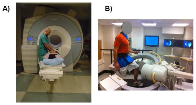
The technique used in this study has been used extensively to investigate knee joint kinematics.(Li et al., 2005, Li et al., 2007) First, each knee was scanned in a relaxed extended position using a 3-Tesla MR scanner (MAGNETOM Trio®, Siemens, Erlangen, Germany) and a double-echo water excitation sequence (Figure 1A). The MRI scans were used to generate sagittal plane images (512 × 512 pixels) with a field view of 16 × 16 cm and 1 mm spacing. The images were then imported into solid modeling software (Rhinoceros®, version 4.0, Robert McNeel & Associates, Seattle, WA) and manually digitized to outline the contours of the femur and tibia. These outlines were used to construct three dimensional geometric models of the knee.
Figure 1.
A) Subjects were first MR-scanned to construct a 3D knee model. B) Following this, each subject performed the step-up exercise while the knee was scanned by the dual fluoroscopic imaging system.
Next, the dual fluoroscopic imaging system setup, previously validated for treadmill gait analysis (Li et al., 2009), was used to determine the kinematics of both injured and intact contralateral knees during the a step ascent (Figure 1A). Two thin pressure sensors (force sensor resistor (FSR), Interlink Electronics, Camarillo, CA) were fixed to the bottom of the subject’s shoe, recording the beginning and end of weightbearing on the studied extremity. Laser-positioning devices, attached to the fluoroscopes, helped to align the target knee within the field of view of the fluoroscopes during the activity.
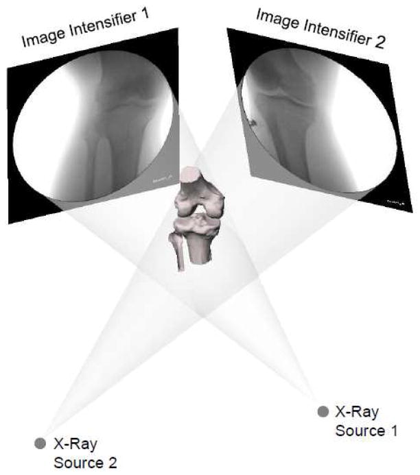
After experiment, the series of fluoroscopic images were imported into the modeling software and placed in calibrated planes to reproduce the orientation of the fluoroscopes during the testing. The 3D MR-based knee model was also imported into the software and manipulated in 6DOF until the projections of the bony model matched the outlined silhouettes of the bones captured on the selected pairs of fluoroscopic images (Figure 2). This process was repeated at each 10% of the activity from the beginning to the end of weightbearing. A previously established and validated coordinate system was used to measure the kinematics.(Defrate et al., 2006)
Figure 2.
Virtual reproduction of the fluoroscopic setup and tibiofemoral kinematics. The 3D MR-based models of the femur and tibia were matched to their projections on the fluoroscopic images.
Statistical Analysis
A two-way repeated measures ANOVA was used to compare the tibiofemoral kinematics of the ACL-injured and ACL-intact knees. The kinematics were the dependent variables and the injury status and time were the independent variables. Level of statistical significance was set at p<0.05. When a statistically significant difference was detected, a post hoc Newman-Keuls test was performed, and the level of significance was again set at p<0.05. The statistical analysis was performed using commercially available software (Statistica® v. 8.0, Statsoft, Tulsa, OK).
RESULTS
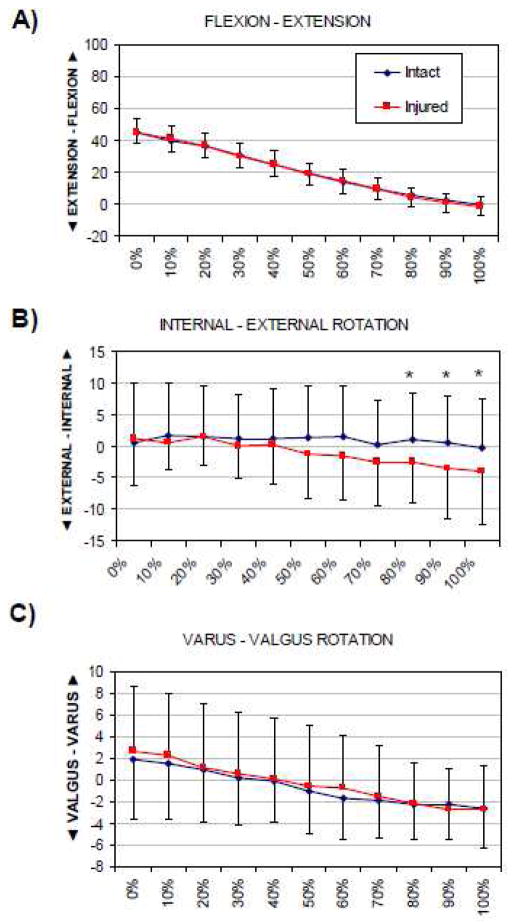
The primary rotation occurred in the sagittal plane (flexion-extension) and its range was on average about 45°. The secondary rotat ions in other rotational planes were of much smaller amplitude (on average <5°). From the beginning to the end of the step ascent the flexion angle consistently decreased from an average of 45° at 0% to an average of 0.2° of hyper extension at 100% of activity progress. There was no significant difference in kinematics in the sagittal plane between the injured and control knees. Significant difference in rotational motion between the two knee conditions was observed in the transverse rotational plane (internal-external rotation) towards the end of the step-up (knee extension). From the initiation of weightbearing until full extension the ACL-injured knee displayed an average of 5° of external tibial rotation. During the last 30% of the activity, this was significantly different from the ACL-intact knee which maintained a relatively constant axial rotation throughout the cycle (p<0.05). Coronal plane rotation was not found to be significantly different between the two conditions at any point during the step-up (p>0.75).
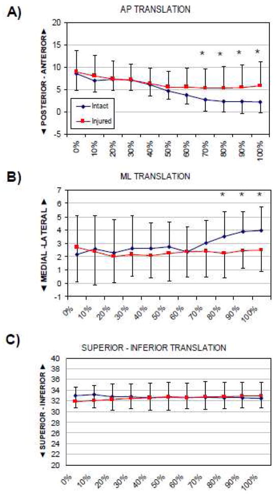
With regards to translational motion, the greatest difference between the ACL-intact and injured knee was found, as expected, in the anteroposterior direction when the knee approached full extension towards the end of the step-up. The ACL-injured knee demonstrated on average 2.5 mm greater anterior tibial shift during the last 40% of the cycle (p<0.01). In addition, significant difference was found in motion in the mediolateral direction. The tibia of the ACL-deficient knee tended to shift more medially as the knee approached full extension. During the last 30% of the cycle this pattern of motion was significantly different from that of the uninjured contralateral knee (p <0.01).
DISCUSSION
This study investigated tibiofemoral kinematics during step-up exercise in patients with ACL deficient knees and uninjured contralateral knees using MRI, dynamic dual fluoroscopy and computer modeling. The data confirmed the initial hypothesis, as it was found that ACL deficiency alters the tibiofemoral kinematics by causing increased anterior tibial translation, medial tibial translation and external tibial rotation. However, these kinematic disturbances were only present towards the end of the step-up as the knee joint approached full extension. The most striking difference between the kinematics of the ACL-injured knee and the contralateral uninjured control was in anteroposterior translation. On average, the tibia of the ACL-deficient knee tended to shift more anteriorly towards the end of the step-up. There are few reports in the literature on the anteroposterior motion in ACL-deficient tibiofemoral joint during step-up or stair climbing. Ahmed et al.(Ahmed and McLean, 2002) studied simulated stair climbing in cadaveric specimens and reported increased anteroposterior translation in ACL deficient knees. The difference was significant only during the terminal stance phase, which is similar to our findings. Vergis et al.(Vergis and Gillquist, 1998), on the other hand, did not find significant difference when they studied the effect of ACL deficiency on sagittal plane knee kinematics during stair climbing using electro-goniometric system. The authors explained that, in those patients, no significant difference was present because they had developed a compensatory mechanism through the action of muscular co-contraction that substituted for the lost ACL. However, those conclusions might have been due to insufficient statistical power. Furthermore, their device was set to zero at full extension of the ACL-deficient knee. Therefore, their data may have accounted for the change in kinematics at full extension of the ACL-deficient knee. As a result, their results might not have found an additional change during weightbearing.
ACL deficiency has also been shown to disturb the flexion-extension motion during stair climbing. Vergis et al.(Vergis and Gillquist, 1998) reported smaller flexion angles and moments for the ACL-deficient knees. They found that maximal anteroposterior translation in the ACL-deficient knees occurred at smaller flexion angles than in the uninjured knees. In contrast to their study, in our study, we did not observe significant differences in flexion between the injured and intact contralateral knee. The discrepancies between these results can be attributed to the different control groups, methodologies and possibly also to presence/absence of gait adaptation mechanisms of the studied subjects. Based on the more recent literature reports, the preferred exercises in the initial stages of rehabilitation after ACL injury and reconstruction are by many considered to be those which employ closed kinetic chain e.g. step-up. Closed chain exercises have been shown to produce smaller anterior displacement of the tibia and smaller anteroposterior shear forces than open kinematic chain exercises.(Bynum et al., 1995, Henning et al., 1985, Lutz et al., 1993, O’Connor et al., 1989, Palmitier et al., 1991, Yack et al., 1993) It is well accepted that closed chain exercises lead to more quadriceps/hamstring/gastrocnemius co-activation that dynamically stabilizes the knee and thus they are believed to be less harmful to the injured knee.(Fitzgerald, 1997, Lutz et al., 1993, Wilk et al., 1996) In contrast to the reports of no significant difference in anteroposterior translation between the ACL-deficient and uninjured knees during step-up exercise or stair climbing(Vergis and Gillquist, 1998), our study found on average 2.5 mm difference in anteroposterior translation between the two conditions during step-up as the knee approached full extension. Concurrently, we found the tibia to shift more medially (~ 1 mm) and externally rotate (~ 5°) towards the end of the step-up. To put these apparently small numbers in clinical perspective, it has been reported that a posterolateral shift of the femur of such magnitudes can substantially alter contact stress distribution in the cartilage by shifting the peak of contact deformation to areas of thinner cartilage.(Van de Velde et al., 2009) In the medial tibiofemoral compartment the contact stress distributions were found to be altered near the medial intercondylar eminence of the tibia - a location where osteoarthritic changes often develop in knees with long-standing ACL insufficiency (Fairclough et al.). The findings of this study question the long-held contention that the step-up exercise is biomechanically safe as it does not alter tibiofemoral kinematics. In this context it is of note that some insurers currently require a minimum 6-month trial of intensified physical therapy before allowing the patients and the surgeons to proceed with ACL reconstruction. Further research is needed to determine whether exercise such as the step-up in the setting of ACL deficiency do not introduce repetitive microtrauma to the cartilage with potentially deleterious consequences.
The results of this study should be interpreted in the context of its potential limitations. First, not all patients included in this study had isolated ACL tears, as the majority of patients have some associated injuries. However, we excluded patients that had insufficiency of other collateral or cruciate ligaments, fracture, required reconstruction other than that of the ACL or removal of more than one third of the meniscus. Next, we did not record the ground reaction forces and instead used the sensors attached to the bottom of the shoe in order to record the start of weightbearing on the studied extremity. Another limitation is that no baseline measure of the change in ACL-deficient knee kinematics was made of the unloaded knee prior to weightbearing. This was not done, as the scope of this investigation was to compare the kinematics of ACL intact and deficient knee during the same loading condition – the step-up exercise. Finally, we only measured kinematics during one cycle of step-up in order to minimize the radiation exposure from the fluoroscopes. However, the step-up exercise has been shown to have reasonable reproducibility.(Yu et al., 1997)
In summary, this study investigated the tibiofemoral kinematics in knees with ACL deficiency during step-up exercise - one of the most commonly performed exercises during physical therapy. Advanced imaging and computer modeling techniques were used to test the hypothesis that ACL deficiency alters tibiofemoral kinematics during the step-up exercise. The data analysis confirmed the initial hypothesis as it was found that ACL deficient knees demonstrated significantly increased anterior tibial translation, medial tibial translation and external tibial rotation towards the end of the step-up as the knee approached full extension. Intensive rehabilitation utilizing the step-up exercise in the setting of ACL deficiency can therefore potentially introduce repetitive microtrauma by way of altered kinematics.
Figure 3.
Tibiofemoral kinematics (rotations) of healthy and ACL deficient knees during the step-up exercise. The values represent motion of the tibia with respect to the femur. Asterisk denotes statistically significant difference at p<0.05.
Figure 4.
Tibiofemoral kinematics (translations) of healthy and ACL deficient knees during the step-up exercise. The values represent motion of the tibia with respect to the femur. Asterisk denotes statistically significant difference at p<0.05.
Acknowledgments
Funding: NIH grants R-01 AR055612 and F-32 AR056451.
This investigation was made possible through the support of the National Institutes of Health (NIH), grants R-01 AR055612 and F-32 AR056451.
Footnotes
Approval by the authors’ institutional review board (IRB) was obtained.
No conflict of interest declared.
Publisher's Disclaimer: This is a PDF file of an unedited manuscript that has been accepted for publication. As a service to our customers we are providing this early version of the manuscript. The manuscript will undergo copyediting, typesetting, and review of the resulting proof before it is published in its final citable form. Please note that during the production process errors may be discovered which could affect the content, and all legal disclaimers that apply to the journal pertain.
References
- AHMED AM, MCLEAN C. In vitro measurement of the restraining role of the anterior cruciate ligament during walking and stair ascent. J Biomech Eng. 2002;124:768–79. doi: 10.1115/1.1504100. [DOI] [PubMed] [Google Scholar]
- BARATTA R, SOLOMONOW M, ZHOU BH, LETSON D, CHUINARD R, D’AMBROSIA R. Muscular coactivation. The role of the antagonist musculature in maintaining knee stability. Am J Sports Med. 1988;16:113–22. doi: 10.1177/036354658801600205. [DOI] [PubMed] [Google Scholar]
- BERCHUCK M, ANDRIACCHI TP, BACH BR, REIDER B. Gait adaptations by patients who have a deficient anterior cruciate ligament. J Bone Joint Surg Am. 1990;72:871–7. [PubMed] [Google Scholar]
- BYNUM EB, BARRACK RL, ALEXANDER AH. Open versus closed chain kinetic exercises after anterior cruciate ligament reconstruction. A prospective randomized study. Am J Sports Med. 1995;23:401–6. doi: 10.1177/036354659502300405. [DOI] [PubMed] [Google Scholar]
- DEFRATE LE, PAPANNAGARI R, GILL TJ, MOSES JM, PATHARE NP, LI G. The 6 degrees of freedom kinematics of the knee after anterior cruciate ligament deficiency: an in vivo imaging analysis. Am J Sports Med. 2006;34:1240–6. doi: 10.1177/0363546506287299. [DOI] [PubMed] [Google Scholar]
- DRAGANICH LF, JAEGER RJ, KRALJ AR. Coactivation of the hamstrings and quadriceps during extension of the knee. J Bone Joint Surg Am. 1989;71:1075–81. [PubMed] [Google Scholar]
- FAIRCLOUGH JA, GRAHAM GP, DENT CM. Radiological sign of chronic anterior cruciate ligament deficiency. Injury. 1990;21:401–2. doi: 10.1016/0020-1383(90)90130-m. [DOI] [PubMed] [Google Scholar]
- FITZGERALD GK. Open versus closed kinetic chain exercise: issues in rehabilitation after anterior cruciate ligament reconstructive surgery. Phys Ther. 1997;77:1747–54. doi: 10.1093/ptj/77.12.1747. [DOI] [PubMed] [Google Scholar]
- HENNING CE, LYNCH MA, GLICK KR., JR An in vivo strain gage study of elongation of the anterior cruciate ligament. Am J Sports Med. 1985;13:22–6. doi: 10.1177/036354658501300104. [DOI] [PubMed] [Google Scholar]
- HOOPER DM, MORRISSEY MC, DRECHSLER W, MORRISSEY D, KING J. Open and closed kinetic chain exercises in the early period after anterior cruciate ligament reconstruction. Improvements in level walking, stair ascent, and stair descent. Am J Sports Med. 2001;29:167–74. doi: 10.1177/03635465010290020901. [DOI] [PubMed] [Google Scholar]
- KELLIS E, BALTZOPOULOS V. The effects of the antagonist muscle force on intersegmental loading during isokinetic efforts of the knee extensors. J Biomech. 1999;32:19–25. doi: 10.1016/s0021-9290(98)00131-6. [DOI] [PubMed] [Google Scholar]
- KOZANEK M, HOSSEINI A, LIU F, VAN DE VELDE SK, GILL TJ, RUBASH HE, LI G. Tibiofemoral kinematics and condylar motion during the stance phase of gait. J Biomech. 2009;42:1877–84. doi: 10.1016/j.jbiomech.2009.05.003. [DOI] [PMC free article] [PubMed] [Google Scholar]
- KVIST J, GILLQUIST J. Sagittal plane knee translation and electromyographic activity during closed and open kinetic chain exercises in anterior cruciate ligament-deficient patients and control subjects. Am J Sports Med. 2001;29:72–82. doi: 10.1177/03635465010290011701. [DOI] [PubMed] [Google Scholar]
- KVIST J, GOOD L, TAGESSON S. Changes in knee motion pattern after anterior cruciate ligament injury - case report. Clin Biomech (Bristol, Avon) 2007;22:551–6. doi: 10.1016/j.clinbiomech.2007.01.003. [DOI] [PubMed] [Google Scholar]
- LI G, DEFRATE LE, RUBASH HE, GILL TJ. In vivo kinematics of the ACL during weight-bearing knee flexion. J Orthop Res. 2005;23:340–4. doi: 10.1016/j.orthres.2004.08.006. [DOI] [PubMed] [Google Scholar]
- LI G, KOZANEK M, HOSSEINI A, LIU F, VAN DE VELDE SK, RUBASH HE. New fluoroscopic imaging technique for investigation of 6DOF knee kinematics during treadmill gait. J Orthop Surg Res. 2009;4:6. doi: 10.1186/1749-799X-4-6. [DOI] [PMC free article] [PubMed] [Google Scholar]
- LI G, PAPANNAGARI R, DEFRATE LE, YOO JD, PARK SE, GILL TJ. The effects of ACL deficiency on mediolateral translation and varus-valgus rotation. Acta Orthop. 2007;78:355–60. doi: 10.1080/17453670710013924. [DOI] [PubMed] [Google Scholar]
- LI G, VAN DE VELDE SK, BINGHAM JT. Validation of a non-invasive fluoroscopic imaging technique for the measurement of dynamic knee joint motion. J Biomech. 2008;41:1616–22. doi: 10.1016/j.jbiomech.2008.01.034. [DOI] [PubMed] [Google Scholar]
- LUTZ GE, PALMITIER RA, AN KN, CHAO EY. Comparison of tibiofemoral joint forces during open-kinetic-chain and closed-kinetic-chain exercises. J Bone Joint Surg Am. 1993;75:732–9. doi: 10.2106/00004623-199305000-00014. [DOI] [PubMed] [Google Scholar]
- O’CONNOR JJ, SHERCLIFF TL, BIDEN E, GOODFELLOW JW. The geometry of the knee in the sagittal plane. Proc Inst Mech Eng H. 1989;203:223–33. doi: 10.1243/PIME_PROC_1989_203_043_01. [DOI] [PubMed] [Google Scholar]
- OSTERNIG LR, CASTER BL, JAMES CR. Contralateral hamstring (biceps femoris) coactivation patterns and anterior cruciate ligament dysfunction. Med Sci Sports Exerc. 1995;27:805–8. [PubMed] [Google Scholar]
- PALMITIER RA, AN KN, SCOTT SG, CHAO EY. Kinetic chain exercise in knee rehabilitation. Sports Med. 1991;11:402–13. doi: 10.2165/00007256-199111060-00005. [DOI] [PubMed] [Google Scholar]
- PATEL RR, HURWITZ DE, BUSH-JOSEPH CA, BACH BR, JR, ANDRIACCHI TP. Comparison of clinical and dynamic knee function in patients with anterior cruciate ligament deficiency. Am J Sports Med. 2003;31:68–74. doi: 10.1177/03635465030310012301. [DOI] [PubMed] [Google Scholar]
- SOLOMONOW M, BARATTA R, ZHOU BH, SHOJI H, BOSE W, BECK C, D’AMBROSIA R. The synergistic action of the anterior cruciate ligament and thigh muscles in maintaining joint stability. Am J Sports Med. 1987;15:207–13. doi: 10.1177/036354658701500302. [DOI] [PubMed] [Google Scholar]
- VAN DE VELDE SK, BINGHAM JT, HOSSEINI A, KOZANEK M, DEFRATE LE, GILL TJ, LI G. Increased tibiofemoral cartilage contact deformation in patients with anterior cruciate ligament deficiency. Arthritis Rheum. 2009;60:3693–702. doi: 10.1002/art.24965. [DOI] [PMC free article] [PubMed] [Google Scholar]
- VERGIS A, GILLQUIST J. Sagittal plane translation of the knee during stair walking. Comparison of healthy and anterior cruciate ligament--deficient subjects. Am J Sports Med. 1998;26:841–6. doi: 10.1177/03635465980260061801. [DOI] [PubMed] [Google Scholar]
- WILK KE, ESCAMILLA RF, FLEISIG GS, BARRENTINE SW, ANDREWS JR, BOYD ML. A comparison of tibiofemoral joint forces and electromyographic activity during open and closed kinetic chain exercises. Am J Sports Med. 1996;24:518–27. doi: 10.1177/036354659602400418. [DOI] [PubMed] [Google Scholar]
- YACK HJ, COLLINS CE, WHIELDON TJ. Comparison of closed and open kinetic chain exercise in the anterior cruciate ligament-deficient knee. Am J Sports Med. 1993;21:49–54. doi: 10.1177/036354659302100109. [DOI] [PubMed] [Google Scholar]
- YU B, KIENBACHER T, GROWNEY ES, JOHNSON ME, AN KN. Reproducibility of the kinematics and kinetics of the lower extremity during normal stair-climbing. J Orthop Res. 1997;15:348–52. doi: 10.1002/jor.1100150306. [DOI] [PubMed] [Google Scholar]


