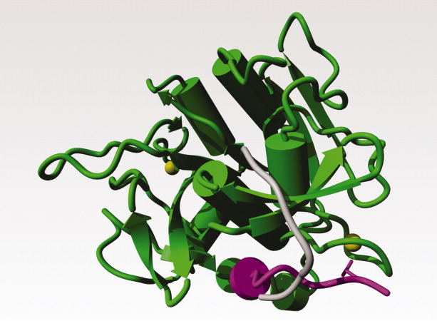
Figure 6.

Subtilisin E from Bacillus subtilis; PDB file 1scj52. The two calcium ions are shown as yellow balls. Residues 1–10 in the mature protease are shown in magenta. Residues 1–10 after the energy minimisation-supported manual operation that brought the N-terminal residue close to the serine in the active site cleft are shown in gray. The side chain of Gln-2, which interacts strongly with one of the calcium ions, is shown as a magenta stick-model.
