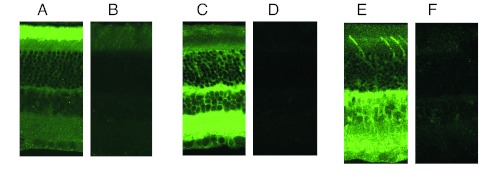
Figure 5.

Immunohistochemical localization of Gβ subtypes in rat retina. (A) FITC localization of Gβ1 heavily stains the outer segment of photoreceptors, with weak staining elsewhere. (C) Gβ2 stains heavily in the outer plexiform layer and inner retina. (E) Gβ3 localizes in the cone outer segment, outer plexiform layer, and selectively stained bipolar cells, amacrine cells and ganglion cell bodies. Specificity of staining was demonstrated by preincubating primary Ab with 10-fold excess of blocking peptides (shown in B, D, and F).
