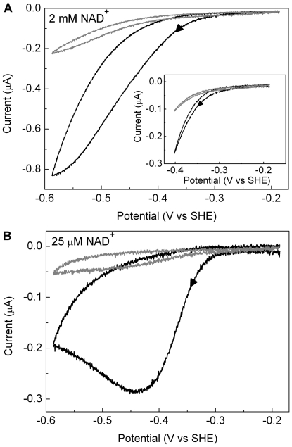
Figure 7. Cyclic voltammograms showing irreversible inactivation of HoxFU at low potentials at different NAD+ concentrations.
In each case the scan rate was 1 mV/s, the electrode was rotated at 2500 rpm, and other conditions were: 50 mM Tris-HCl buffer, pH 8.0, 30°C; (A) 2 mM NAD+ and (B) 25 µM NAD+. The first cycle for each film is shown as a thick line and the second cycle is shown as a thin line. Arrows indicate the direction of scan. Inset in (A) shows a scan over a narrower potential range.

