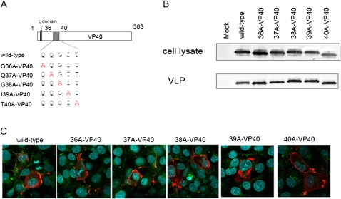
Figure 4.
Single alanine-scanning of VP40 at amino acid residues 36–40. A, Schematic diagrams of alanine-scanning mutants. B, Comparison of virus-like particle (VLP) budding induced by the alanine-scanning mutants. We transfected 293T cells with a plasmid expressing each mutant. At 48 hours after transfection, the VLP budding assay was performed. All mutants produced VLPs in the supernatant as effectively as wild-type VP40. C, Intracellular localization of single-alanine mutant 293 cells were transfected with a plasmid expressing a cMyc-tagged VP40 mutant. At 24 hours after transfection, cells were stained with an anti-cMyc antibody (red) and an anti-CD63 antibody (green). Nuclei were stained with Hoechst (Invitrogen) (blue). The intracellular localizations of I39A-VP40 and T40A-VP40 were different from that of the wild type.

