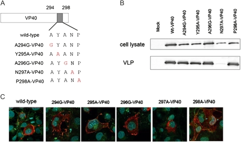
Figure 6.
Virus-like particle (VLP) budding induced by the single-alanine mutants. A, Schematic diagram of the single-alanine mutants. B, Comparison of VLP budding induced by the alanine mutants. We transfected 293T cells with a plasmid expressing each mutant. At 48 hours after transfection, the VLP budding assay was performed. The intensity of the VP40 bands was quantified in relation to the amount of VP40 in lane 1, which was set to 100%. The result presented is the mean of 3 independent experiments. The level of VLP budding for N297A-VP40 was decreased to ∼20% of that of the wild type. C, Intracellular localization of single-alanine mutants. We transfected 293 cells with a plasmid expressing a cMyc-tagged VP40 mutant. At 24 hours after transfection, cells were stained with an anti-cMyc antibody (red) and an anti-CD63 antibody (green). Nuclei were stained with Hoechst (Invitrogen) (blue). The intracellular localization of N297A-VP40 was different from that of the wild type.

