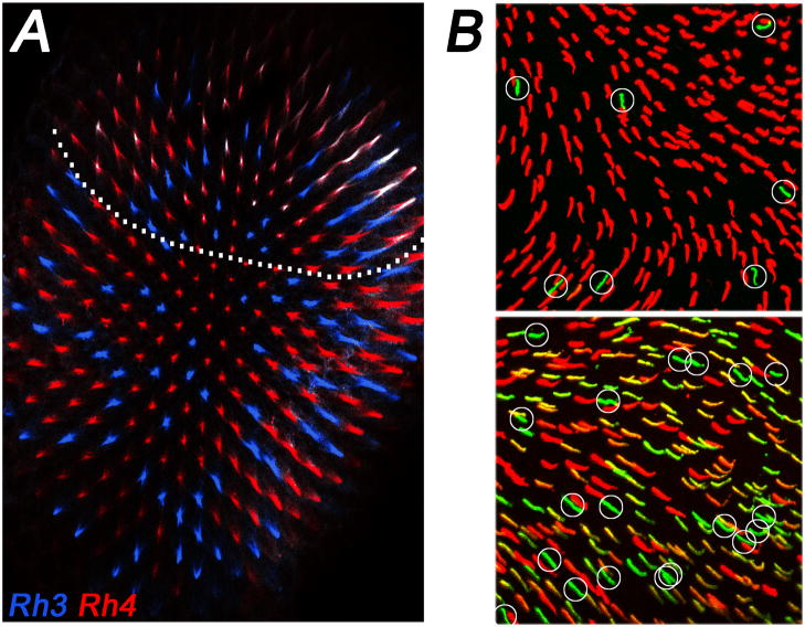
Figure 3.
Regional differences in opsin expression. (A) Fly dorsal yellow ommatidia coexpress (white) Rh3 (blue) and Rh4 (red). Note that these specialized ommatidia occur only in the dorsal third of the eye (indicated by dotted line). (B) The dorsal mouse retina (top) has mostly M opsin (red) and few S opsin (green, circled) positive cones. In the ventral retina (bottom), the majority of cones show variable levels of coexpression of M and S opsin. Cones that appear to predominantly express S opsin (green, circled) are much more frequent than in the dorsal part of the retina. Figure adapted from (Haverkamp et al., 2005).

