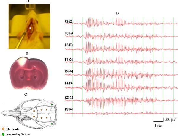
Figure 1.

(A) Placement of an argon laser beam on the exposed skull of a rat. The argon laser beam is used to create the infarct in the lesioned groups of animals. (B) A cortical infarct generated by laser stimulation as assessed by TTC staining. (C) Placement of recording and anchoring screws on the rat's skull. (D) Sample of a generalized SWD recorded from a Fischer 344 rat.
