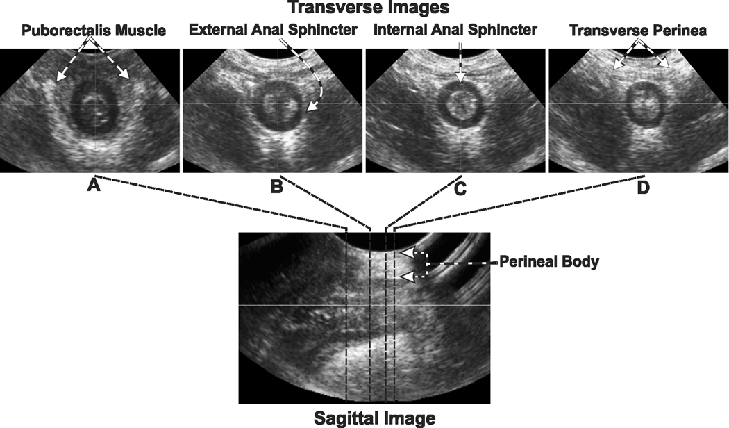
Figure 4. Transverse & Sagittal Image of the Anal Canal.
Lower panel shows the ultrasound image of the anal canal in the mid-sagittal plane. The images in the upper row show transverse or tomographic axial images at different levels along the cranio-caudal extent of the anal canal. Note the appearance and location of internal anal sphincter (IAS), external anal sphincter (EAS) and puborectalis muscle (PRM) along the length of the anal canal. Also note that the inner fibers of the EAS surround the whole circumference of the anal canal but the outer fibers merge into the right and left transverse perinei muscles anteriorly.

