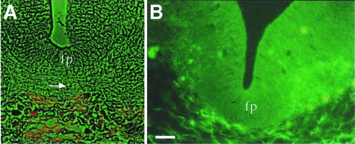
Figure 1.

Immunolocalization of F-spondin protein in E13 rat embryo. Sections were labeled with the anti-reespo domain Ab R5 (15) [tetramethylrhodamine B isothiocyanate (TRITC) in A and FITC in B]. (A) Cross section at the spinal cord level. F-spondin protein is localized in the basal membrane that underlies the floor plate (fp). Note that the crossing fibers of the commissural axons (arrow) are in contact with the F-spondin protein. (B) Cross section at the midbrain level. F-spondin protein is localized at the extracellular matrix that underlies the brain, as well as in the neuroepithelium lateral to the floor plate. (Scale bar = 25 μm; A is at the same magnification.)
