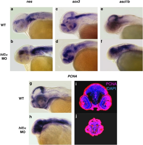
Figure 3.
The surviving neural progenitor cells in hif2α morphants continue to proliferate without terminally differentiating. (a–f) Lateral views of nes (a and b), sox3 (c and d) and ascl1b (e and f) expression in WT (a, c and e) and hif2α ATG-MO (b, d and f) 48 h.p.f. embryos. nes, sox3 and ascl1b were highly expressed in the NPCs but not in mature neural cells. The high level of nes, sox3 and ascl1b transcriptions in hif2α morphants suggests that the NPCs did not differentiate into mature neural cells. (g and h) Lateral views of pcna transcription in WT (g) and hif2α ATG-MO (h) 48 h.p.f. embryos. (i and j) Immunofluorescent staining of PCNA (in red) in transverse brain sections of WT (i) and hif2α ATG-MO (j) 48 h.p.f. embryos. Nuclei are labeled by DAPI staining (in blue). The increased pcna expression in the CNS of hif2α MO embryos indicates that the NPCs were proliferating

