
Figure 2.
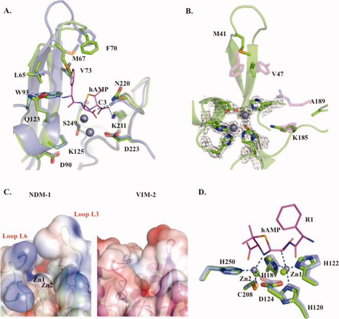
Apo-NDM-1/hydrolyzed ampicillin (hAMP)-bound NDM-1 and apo-NDM-1/apo-VIM-2 active-site comparisons. A: Apo-NDM-1/hAMP NDM-1 active-site overlay: apo-NDM-1 backbone (green), hydrolyzed ampicillin (hAMP) product complex (gray, PDB ID: 3Q6X)13 with selected active-site residues (sticks) labeled.13 B: Apo-NDM-1/apo-VIM-2 active-site overlay: apo-NDM-1 backbone (green), selected NDM-1 and VIM-2 active-site residues are shown as green and magenta sticks with cpk atom coloring, respectively. The 2Fo − Fc map at 2σ is shown for zinc ions and coordinating ligands. C: Electrostatic surface representation of apo-NDM-1 and apo-VIM-2 active-site clefts, calculated using APBS software, with a PARSE force field contoured at −3 and +3. D: Zinc coordination: zinc ions and ligands are in green and gray for apo-NDM-1 and hAMP-bound NDM-1. Coordination interactions are blue dashes, and hAMP is in cpk colored stick representation (carbons are magenta).