
Figure 8.
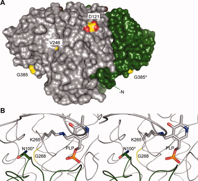
Hereditary sensory and autonomic neuropathy type 1 (HSAN I)–linked mutations on SpSPT. A: Surface representation of holo-SpSPT (PDB code 2JG2117). Subunit A is colored pale gray, whereas subunit B is dark green. The first 20 residues of subunit B are represented by a green dashed line. The residues of SpSPT corresponding to mutations linked to HSAN I are colored yellow and atom color. The bound Mg ions appear as brown spheres. Compared to Figure 6(A), the dimer was rotated 90° anticlockwise along an axis parallel to the plane of the sheet. This figure is linked to Table I. B: Stereoview of the active site. The residue G268 is colored yellow.