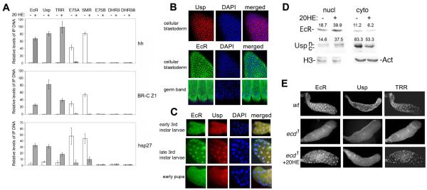
Figure 1. Ecdysone-dependent changes in association with target genes and subcellular distribution of ecdysone receptor components.
(A) Relative amounts of EcR, Usp, TRR, E75A, SMR, E75B, DHR3, and DHR38 at the hh, BR-C Z1, and hsp27 promoters in S2 cells. Bars show means ± standard deviations above background from 3 ChIP experiments. Background was determined by omitting precipitating antibody. Primer set 4 (Fig. 4C) was used for BR-C Z1.
(B, C) Immunostaining with EcR and Usp antibodies of cellular blastoderm and germ band-retracted embryos (B), and salivary glands from early (1 day old), late 3rd instar larvae and early pupae (stages II and III, respectively, Fig. 4A).
(D) Western blots of EcR and Usp in extracts from nuclei and cytoplasm of S2 cells. Nuclear (n) and cytoplasmic (c) forms of Usp are indicated. Actin and histone H3 antibodies served as loading controls for cytoplasmic and nuclear extracts, respectively. Numbers in each column are the quantified band intensities, normalized to loading controls. The change in nuclear/cytoplasmic ratio with addition of 20HE is 3.88 for EcR and 4.02 for Usp.
(E) Salivary glands of wild-type (wt) and homozygous ecd1 3rd instar larvae immunostained with Usp, EcR and TRR antibodies. Wild-type and ecd1 larvae were grown at 29° C, (restrictive temperature), starting at the end of the 2nd larval instar.

