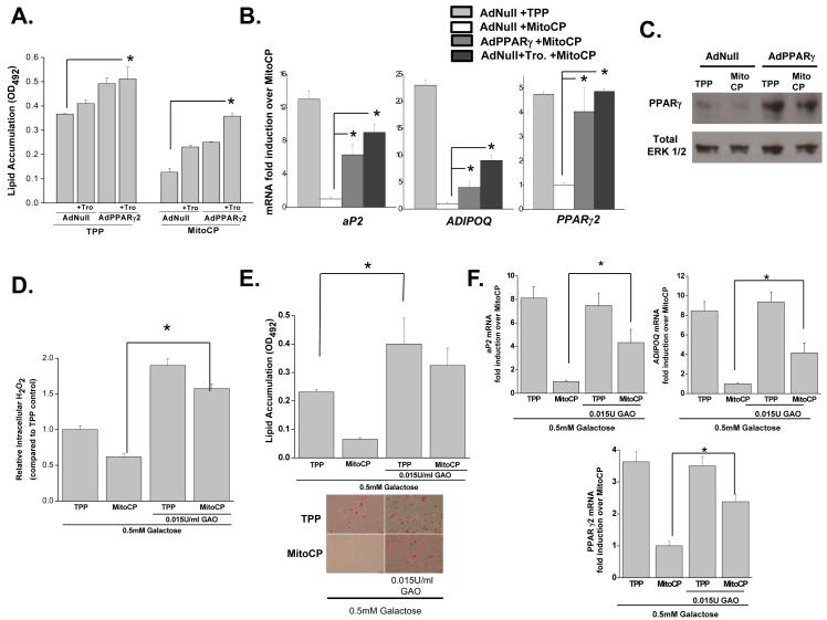
Figure 2. Exogenous PPARγ or H2O2 rescues adipocyte differentiation in the presence of mitochondrial targeted antioxidants.
(A) Human MSCs were infected with AdPPARγ2 or AdNull at day 1 and treated with TPP (500nM) or MitoCP (500nM) +/− 5 μM troglitazone (Tro) starting at day 2. Subsequently, at day 21 of differentiation cells were stained with Oil Red O and optical density (OD) values were assessed. N=3 ± SEM.
(B) Gene expression of PPARγ2 and its target genes at day 7 of differentiation in human MSCs infected with AdPPARγ2 or AdNull at day 1 and treated with TPP (500nM) or MitoCP (500nM) +/− 5 μM troglitazone (Tro.) starting at day 2. N=3 ± SEM.
(C) Western blot analysis of PPARγ protein at Day 5 of adipocyte differentiation. Human MSCs were infected with AdNull or AdPPARγ2 at day 1 and administered 500 nM of MitoCP or TPP control starting at day 2.
(D) Intracellular H2O2 levels of human MSCs treated with galactose (0.5mM) with or without galactose oxidase (GAO, 0.015U/ml) in the presence of 500 nM of TPP or MitoCP starting at Day 2. N=3 ± SEM.
(E) Human MSCs were treated with galactose (0.5mM) with or without GAO (0.015U/ml) plus 500 nM of TPP or MitoCP starting at day 2. Subsequently, at day 21 of differentiation cells were stained with Oil Red O and optical density (OD) values were assessed. N=3 ± SEM.
(F) Human MSCs were treated with galactose (0.5mM) with or without GAO (.015U/ml) plus 500 nM TPP or MitoCP starting at day 2. Gene expression was analyzed at day 7 of differentiation. N=3 ± SEM.

