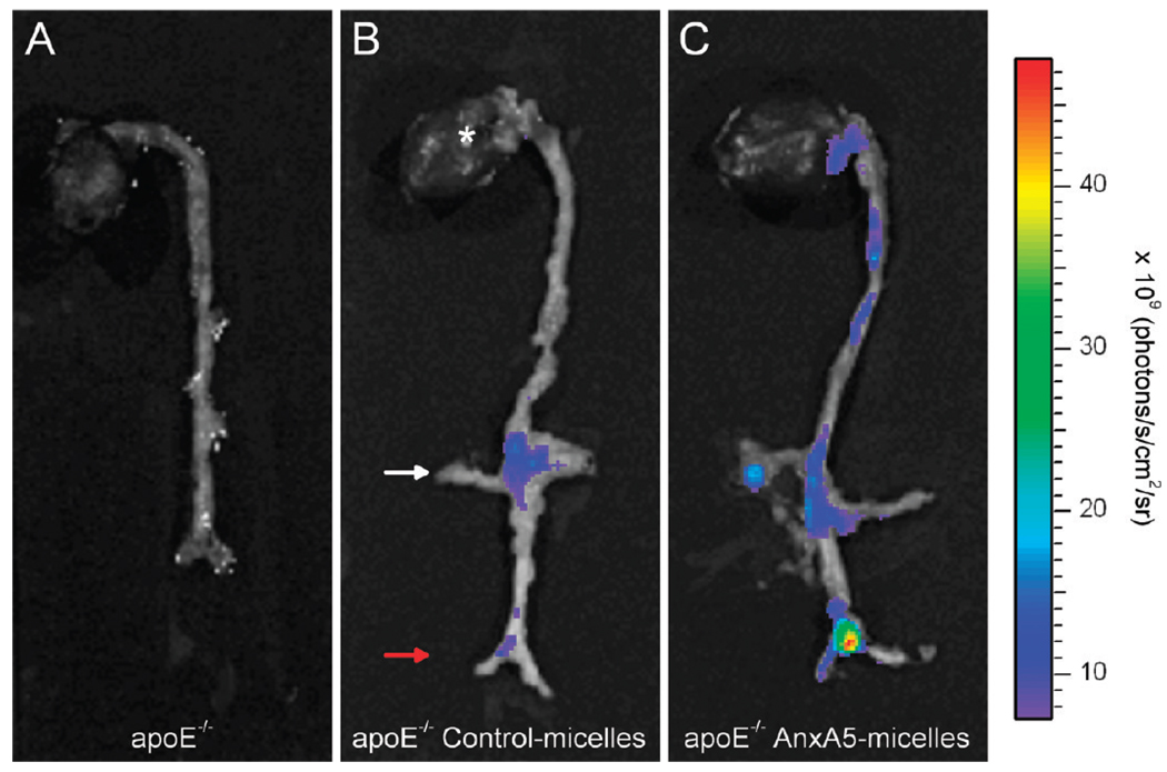
Figure 5.
Ex vivo near-infrared fluorescence imaging (NIRF) of whole aortas from ApoE−/− mice (A) without contrast agent injection and at 24 h after injection of (B) control-micelles and (C) annexin A5-micelles. Micelles contained Cy5.5-PEG2000-DSPE. The orientation of the aorta is indicated by characteristic regions in (B), such as the heart (*), a renal branch (white arrow),and the aortic bifurcation (red arrow). In vivo MR images were acquired in the area between the white and the red arrow.

