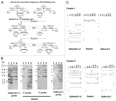
Figure 2.
RFLP analysis of the S. meliloti isolates. (A) Schematic diagrams of intron-less and intron-invaded DNA sites. The DNA probes used for DNA hybridization are indicated below each diagram. Numbers indicate relevant nucleotide positions within the exons and intron sequences. (B) Examples of RFLP analysis depicted in (C) for XhoI-digested total DNA from S. meliloti 1021 (lane 1) and representative isolates from cluster I (B4, lane 2) and cluster II (B10, lane 3 and B1, lane 4), with probes for the mobile elements indicated under each part and represented in (A). DNA molecular size markers are indicated on the left of the first part. (C) Schematic diagrams of Southern blot hybridizations of XhoI-digested total DNA from isolates of clusters I (above) and II (below). The mobile elements used as probes are indicated at the bottom. DNA molecular size markers (λ) are also shown. Asterisks (*) indicate that the band hybridizes only with the probe for the 5′-end of RmInt1.

