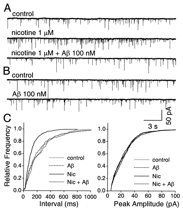
Figure 5.
Aβ1–42 blockade of nicotine-induced increases in spontaneous mEPSC frequency in hippocampal neurons. (A) Whole-cell perforated-patch–clamp recording from a neuron showing that the rate of spontaneous mEPSCs (Top) is increased by application of 1 μM nicotine (Middle) as shown previously (16) and that the nicotine-induced increase can be blocked by 100 nM Aβ1–42 (Aβ; Bottom). Tetrodotoxin and bicucculine were present to block action potentials and GABAA receptors, respectively. (B) Same neuron as in A, showing that Aβ1–42 does not depress the basal rate of spontaneous mEPSCs. (C) Cumulative distribution plots showing that 100 nM Aβ1–42 prevents 1 μM nicotine (Nic) from increasing the frequency of spontaneous mEPSCs but that the peptide has no effect on the basal rate (Left); in contrast, neither nicotine nor Aβ1–42 has any effect on the amplitude distribution of the spontaneous mEPSCs (Right).

