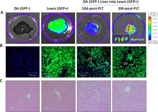
Figure 2. Host repopulation of liver allograft in the animals displaying long term acceptance.
(A) The Xenogen imaging system was used to study DA livers into GFP Lewis hosts with dual drug treatment after partial (50%) liver transplantation. The non-transgenic donor DA liver has no GFP expression. However, the transplanted donor liver graft shows GFP positive at 10 days and a high degree of fluorescence at three months after transplant into a GFP+ Lewis recipient treated with plerixafor and tacrolimus. (B) Liver tissue sections analyzed by fluorescent microscopy. GFP positive cells were present in central vein areas on day 10 after transplantation. Most of the hepatocytes and non-parenchyma cells are GFP positive indicating virtually complete host repopulation_at 3 months after transplantation. Representative photographs of n = 3 individual transplant samples. Images were photographed with a 40× objective. (C) H&E staining showed bile duct regeneration (10 days) and small size hepatocytes in the central vein areas (3 months) suggesting that repopulation and/or remodeling is ongoing. Representative photographs of n = 3 individual transplant samples. Images were photographed with a 20× objective.

