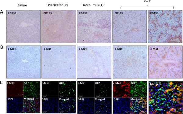
Figure 5. Recruitment of host bone marrow stem cells to rat liver transplants following treatment with dual drug therapy.
Immunohistochemistry staining for CD133 (A) and c-Met (B) in tissue sections from liver allografts at 7 days after transplantation. CD133 or c-Met positive cells are brown. Representative photographs of n = 3 individual transplant samples per group. Images were photographed with a 20× or 40× (right panels) objective. (C) Double fluorescence staining for c-Met and GFP at 7 days after transplantation. Sections stained with both anti-c-Met and anti-GFP antibodies show all c-Met positive cells (red) stained for recipient GFP (green) in rats treated with dual drug therapy. Representative photographs of n = 3 individual transplant samples per group. Images were photographed with a 40× objective.

