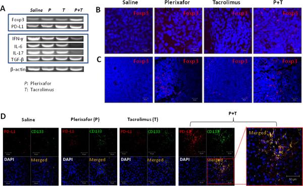
Figure 6. Increased Foxp3 and PD-L1 expressions in transplanted rats treated with dual drug therapy.
(A) Semi-quantitative PCR analysis of liver allografts at 7 days after transplantation. Representative graphs of three individual samples. The mRNA expression of programmed cell death ligand-1 (PD-L1) and Treg marker Foxp3 was significantly increased in the animals receiving dual drug treatment. Interestingly, IFN-γ, IL-17 and IL-6 were all significantly decreased in the dual treatment group, although TGF-β remained unchanged. (B) Detection of Foxp3+ cells in liver allografts and spleens by immunofluorescence staining. Cell nuclei were stained blue with DAPI. The number of Foxp3 positive cells (red) was significantly higher in tissue sections from liver allografts and spleens in the dual treatment group at 7 days after transplantation. (C) Double fluorescence staining for PD-L1 and CD133 at 7 days after transplantation. Sections stained with both anti-PD-L1 and CD133 antibodies show all PD-L1 positive cells (red) stained for CD133 (green) in rats treated with dual drug therapy. Representative photographs of n=3 individual transplant samples per group. Images were photographed with a 40× objective.

