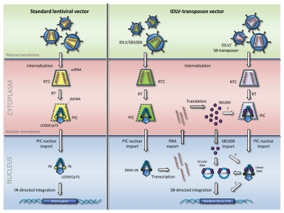
Figure 1.
Schematic representation and comparison of gene delivery mediated by conventional lentiviral vectors (left) and lentivirus-transposon vectors (right). The figure shows the internalization of the nucleocapsid core, shown in yellow, green and purple for the different vectors. Reverse transcription (RT) of vector RNA occurs inside the reverse transcription complex (RTC); after DNA synthesis the increasingly porous complex is referred to as the preintegration complex (PIC). Several cellular proteins become part of the PIC; only LEDGF/p75 is shown. LEDGF/p75 interacts with viral integrase (IN) proteins, shown as yellow circles attached to the terminal regions of the viral DNA. IN proteins harboring the inactivating D64V mutation are indicated by yellow circles marked with a cross. In case of the IDLV-transposon system, cells are transduced with two vectors, one carrying the SB100X expression cassette (green) and one carrying the SB transposon vector (purple). RNA encoding SB100X is produced from episomal viral DNA and exported from the nucleus prior to translation in the cytoplasm. Subunits of SB100X (indicated by purple circles) may be incorporated directly into the PIC (indicated by a stippled arrow and a question mark) or be directly imported into the nucleus where it may interact with both linear DNA and circular DNA intermediates (1- and 2-LTR circles) to facilitate transposition into randomly chosen TA-dinuclotides in the genome. It is not known whether linear viral DNA serves as a substrate for SB transposition. Single-stranded RNA (ssRNA) is shown in red, double-stranded DNA (dsDNA) in blue.

