Fig. 1.
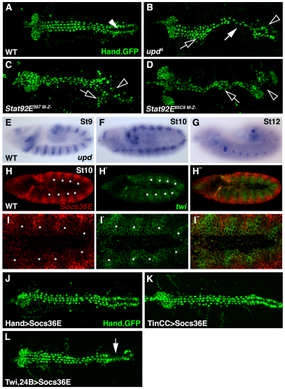
The Jak/Stat pathway regulates cardiac morphology and is active in the dorsal mesoderm during cardiac precursor specification. (A) WT Hand.GFP embryos express nuclear-localized GFP throughout the dorsal vessel. The contractile ‘heart’ (arrowhead) positioned at the posterior of the dorsal vessel pumps hemolymph through the anterior aorta. (B) Zygotic upd4 embryos lose cardioblast cell-cell adhesion (white arrow) causing inappropriate cell aggregations (open arrow) and incomplete heart lumen formation (open arrowhead). (C,D) Embryos maternal and zygotic mutant for Stat92E397 (C) or Stat92E85C9 (D) phenocopy the dorsal vessel defects of upd embryos. (E-G) upd mRNA expression in WT embryos. upd expression in the ventral ectoderm is segmented during St9 (E). upd expression is restricted to the tracheal system and hindgut in St10 (F) and St12 (G) embryos. (H-I′) St10 WT embryo double labeled for Socs36E (red) and twi (green) mRNA. Socs36E and twi expression is complementary in the dorsal mesoderm. Regions of the mesoderm segment expressing high levels of twi (asterisks) express low levels of Socs36E. (J,K) St17 embryos expressing Hand.GFP. Expressing Socs36E in the heart after cardioblast precursor specification using Hand.gal4 (J) or TinCC.gal4 (K) does not affect heart morphology. (L) Expressing Socs36E in the heart prior to cardioblast precursor specification using Twi.gal4, 24B.gal4 induces mild cardioblast cell-cell adhesion defects. A-G show dorsal views of Hand.GFP expression in live St17 embryos. Embryos are oriented with anterior to the left.

