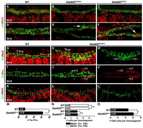
Fig. 3.
JAK/Stat signaling regulates pericardial cell number. (A-F) Lateral views of St13 (A-C) and dorsal views of St16 (D-F) embryos labeled for Mef2 protein and mid mRNA. WT and Stat92EM–Z– embryos express mid in all Mef2+ cardioblasts by St13. mid expression persists to St16 even in the aggregated cardioblasts of Stat92EM–Z– embryos (arrows). (G-H′) Lateral views of St13 embryos labeled for Tin and Mef2. (G) WT St13 embryos express Tin in four cardiomyocytes per heart hemisegment (arrowheads) and in a subset of pericardial cells. Mef2 is not expressed in pericardial cells. (H) Stat92EM–Z– embryos express Tin in four cardiomyocytes in a majority of hemisegments but the number of Tin expressing pericardial cells is significantly increased (open arrows). (I-J′) Dorsal views of St16 embryos labeled for Tin and Prc. (I) Prc localizes to the extracellular matrix between cardiomyocytes and pericardial cells in WT embryos. (J) Ectopic Tin cells positioned lateral to the cardioblasts (open arrowheads) co-express Prc in Stat92EM–Z– embryos. (K-L′) Lateral views of St13 embryos labeled for Odd and Mef2. (K) WT embryos express Odd in four pericardial cells per hemisegment. (L) The number of Odd+ pericardial cells is significantly increased in Stat92EM–Z– embryos. (M) Quantification of Tin+ pericardial cells (PCs) in one half of the mesoderm at St13. Tin+ pericardial cells were identified by lack of Mef2 expression. (N,O) Segmental quantification of Tin+ and Tin–cardioblasts (CBs) at St13 (N) and Odd+ pericardial cells at St13 (O). **P<0.005, ***P<0.001, unpaired t-test. n.s., non-significant. Error bars represent s.e.m. Embryos are oriented with anterior to the left.

