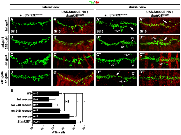
Fig. 4.
JAK/Stat signaling in the mesoderm restricts pericardial cell number. Embryos labeled for HA and Tin. The crossing scheme is detailed in the text. (A-D) St13 embryos lacking the maternal contribution of Stat92E (Stat92EM–) have an increased number of Tin+ cells compared with WT embryos. (A′,B′,D′) Stat92EM embryos that express Stat92E-HA in the mesoderm show WT levels of Tin+ cells. (C′) Stat92EM– embryos that express Stat92E-HA in the ectoderm show a significant increase in the number of Tin+ cells compared with WT embryos. (A′-D′) St16 Stat92EM– embryos show a loss of cardioblast cell-cell adhesion (white arrows), collapsed ‘heart’ (open arrowheads) and inappropriate cell aggregations (open arrows). (A′′-C′′) Stat92EM– embryos that express Stat92E-HA in either the mesoderm or the ectoderm also show these three cardiac phenotypes, although they are less severe. (D′′) Stat92EM– embryos that express Stat92E-HA in the ectoderm and the mesoderm show mostly normal heart morphology. (E) Quantification of Tin+ cells in St13 embryos from the genotypes shown in A-D. *P<0.05, unpaired t-test. NS, non-significant. Error bars represent s.e.m. Embryos are oriented with anterior to the left.

