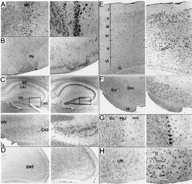
Figure 2.
Distribution of DGKɛ in the mouse brain as measured by in situ hybridization. Adjacent sagital sections were hybridized with sense (Left) or antisense (Right) digoxigenin-labeled probe prepared from a 0.9-kb EcoRI fragment of murine DGKɛ. (A) Staining of the olfactory bulb was most notable in mytrial cells (MC; ×150) and to a lesser extent in the granular cells. (B) Staining was notable in the piriform cortex (Pir; ×60) but was not prevalent in adjacent cortical structures including the insular cortex (data not shown). (C) In the hippocampus (×40, and boxed regions at ×160, Lower), intense signal in the pyramidal cells of CA3 but only weak staining of dentate granular (DG) cells. Pyramidal cells of CA1 were labeled throughout, but no signal in the stratum oriens (so) and only inconsistent staining of cells in other hippocampal regions was observed. (D) Prominent signal for DGKɛ RNA detected in the entorhinal cortex (ENT; ×60) and especially cells of the outer layers. (E) Staining of the medial occipital (neo) cortex (×80) in all layers but was most prominent in the pyramidal cells (pc) of layer 5. Definition of the layers was based upon adjacent sections counterstained with Giemsa (data not shown). No staining of cells over background in the internal capsule (ic) was detected. Staining of cells in the thalamus or in structures of the basal ganglia was occasional or absent, except in F (×60) for the substantia nigra reticulata (Snr). Staining of cells in the substantia nigra compacta (Snc) was present but inconsistent. The cerebral peduncle (cp) is identified. (G) In the cerebellum (×150), staining was most intense in Purkinje cells (Pkj) but could be identified above background in cerebellar granular cells (Gc) and cells of the layer molecular (mol). Staining of cells throughout the hindbrain and Pons was observed including trigeminal nuclei and the superior olive (data not shown). (H) Staining was particularly intense in the lateral reticular nucleus (LNR; ×150).

