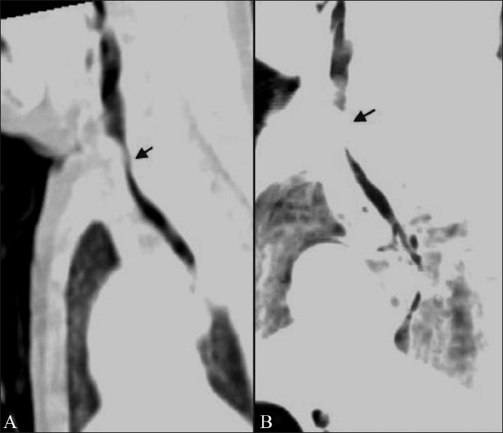
Figure 1 (A, B).

Tracheomalacia. Sagittal MPR (A) and minIP (B) images show a wavy contour of the trachea in this case of tracheomalacia with narrowing (arrow) in its upper portion. The minIP image overestimates the degree of narrowing and the segment length
