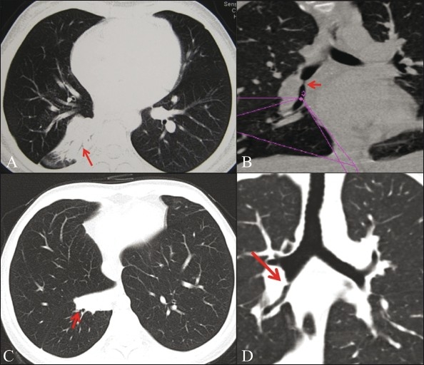
Figure 3.

Post-tuberculous bronchial narrowing. Axial image (A) shows collapse (arrow) of the right lower lobe (RLL). MPR image (B) shows a short segment narrowing (arrow) in the bronchus intermedius. Post-balloon dilatation axial image (C) shows improvement in the RLL aeration (arrow) with residual collapse of the medial basal segment (arrow). Coronal MPR image (D) shows improvement in the caliber of the previously narrowed bronchus intermedius (arrow)
