Figure 4 (A, B).

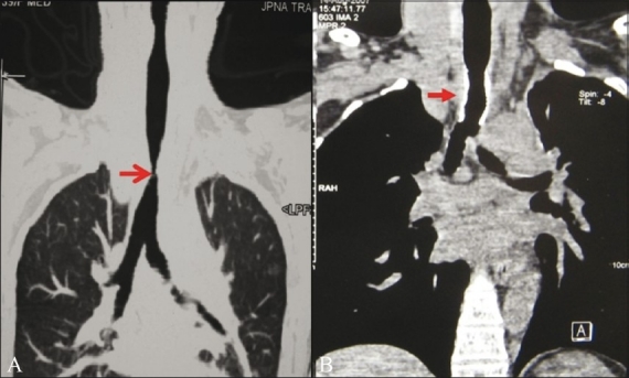
Post-intubation stenosis: Curved coronal lung window MPR image (A) shows segmental narrowing of the trachea (arrow). Post-metallic stenting soft tissue coronal MPR image (B) shows improvement in tracheal caliber (arrow)

Post-intubation stenosis: Curved coronal lung window MPR image (A) shows segmental narrowing of the trachea (arrow). Post-metallic stenting soft tissue coronal MPR image (B) shows improvement in tracheal caliber (arrow)