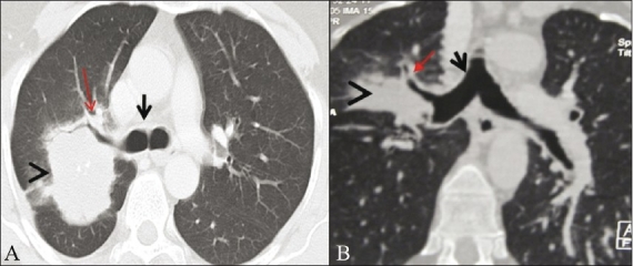
Figure 5 (A, B).

Bronchogenic carcinoma. Axial (A) and coronal MPR (B) lung window images show the presence of a mass in the posterior segment of the right upper lobe (arrowhead), which encases the right upper lobe (RUL) bronchus (arrow). The distance of the mass from the carina (short arrow) the on the axial image (1.5 cm) was less than on the MPR image (3.3 cm)
