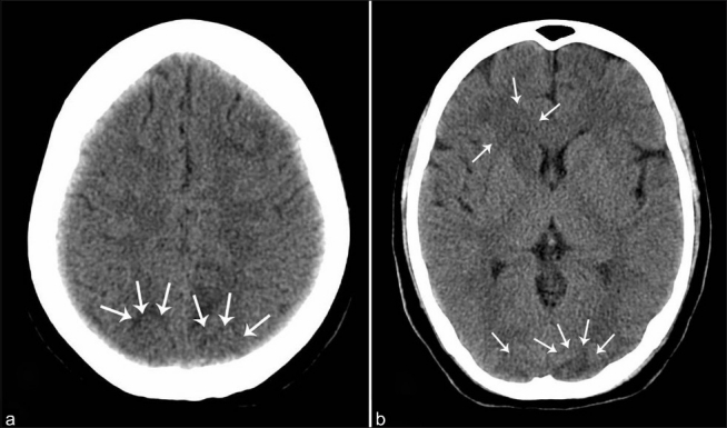
Figure 1.

(a) Plain CT study of brain showing intra-axial hypodensities involving white matter regions of bilateral occipital lobes. (b) Plain CT brain showing intra-axial hypodensities involving white matters of bilateral parieto-occipital lobes and right caudate nucleus
