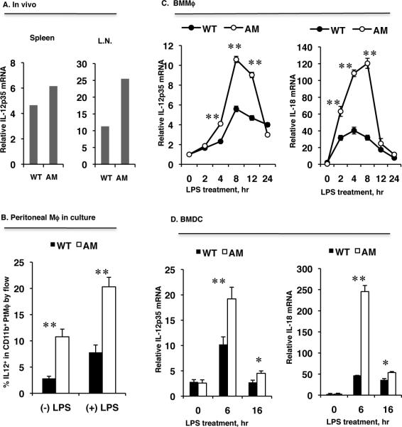
Figure 6. dko DCs and macrophages produce more IL-12 and IL-18 in response to LPS treatment than wild-type cells.
A. IL-12p35 mRNA levels in total splenic and lymph node RNA prepared and pooled from three naïve WT and three dko mice measured by qPCR. B, Thioglycollate-induced peritoneal macrophages (Mϕ) were treated with LPS for 4 h, then IL-12 expression on the CD11b-gated macrophages was analyzed by FACS. The results are the mean ± SD for n=3 mice. C and D, IL-12p35 and IL-18 mRNA levels in bone marrow-derived macrophages (BMMϕ) (C) or bone marrow-derived DCs (BMDC) (D) during LPS treatment measured by qPCR. The results are the mean ±SD for 3 wells per group in one experiment and are representative of those obtained in three experiments **p<0.05, **p<0.001 by the one-way ANOVA test using ProStat Ver 5.5.

