
Fig. 9.
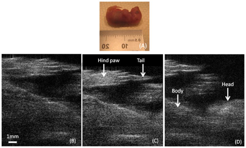
Images of excised mouser embryo (E18.5). Transudcer is positioned along the body of the embryo. (A) Picture of excised mouse embryo. (B) Bottom part of the embyro. (C) Middle part of the embyro(abdomen and thorax). (D) Top part (thorax and head).. The dynamic range is 50dB.