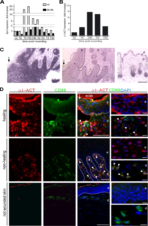
FIGURE 1.
Spi-2/α1-ACT is expressed during the acute phase response post skin injury. A, quantitative RT-PCR analysis of RNA from wound tissue at indicated time points after injury revealed a dramatic increase in Spi-2 mRNA in wild-type (wt) mice (wounds per time point n = 4), and at all time points expression in wounds of db/db mice (wounds per time point n = 4) was attenuated. B, levels of α1-ACT mRNA were also strongly up-regulated in normal healing human wounds (wounds per time point n = 4); ns, not wounded skin (n = 4); h, hours; d, day. C, paraffin sections of normal healing wounds (day 5 post-injury) (left and middle) and intact skin (right) were hybridized to a digoxigenin-labeled antisense probe for Spi-2 mRNA (left and right) and sense probe; reaction product appears in purple; some sections were counterstained with nuclear fast red. D, double immunofluorescence staining of α1-ACT (red) and CD68 (green) in tissue of a healing wound (day 3 post-injury), a nonhealing human wound, and not wounded skin; DAPI counterstaining of nuclei (blue); dotted line indicates basement membrane; arrow indicates wound edge; arrowheads indicate double-positive cells. e, epidermis; d, dermis. Scale bars, 100 μm in C and D; 20 μm in D, right panel.

