FIGURE 2.
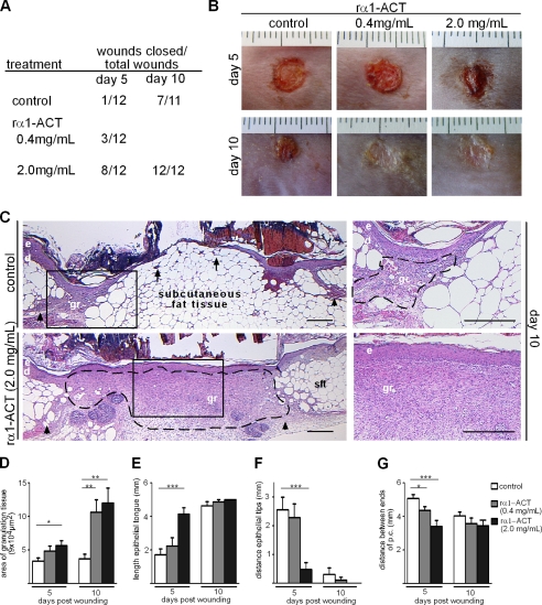
Topical application of rα1-ACT accelerates granulation tissue formation and epithelialization in diabetic mice. Repetitive application of rα1-ACT (concentrations as indicated) significantly accelerated wound closure kinetics of db/db mice compared with vehicle-treated controls. A, presented are numbers of closed wounds versus total number of wounds for indicated conditions and time points post-injury. B, representative macroscopic appearance of wounds at indicated time points post-injury; day 5 post-injury, wounds treated with 2.0 mg/ml rα1-ACT revealed almost complete epithelialization; day 10 post-injury, almost all wounds treated with both concentrations are closed. C, representative H&E staining of wound tissues post-injury; in rα1-ACT treated wounds a highly vascularized and cellular granulation tissue developed that is covered by a hyper-thickened and closed epithelium; in contrast, in control mice scarce granulation tissue developed at wound edges and a thin, not closed, epithelium overlays the massive subcutaneous fat layer; right panel depicts magnifications of rectangles in left panel. D–G, morphometric analysis of wound tissue at different time points post-injury. D, area of granulation tissue (p = 0.01, control versus 2.0 mg/ml rα1-ACT day 5; p = 0.004, control versus 0.4 mg/ml rα1-ACT day 10; p = 0.003, control versus 2.0 mg/ml rα1-ACT day 10). E, length of epithelial tongue (p = 0.0001, control versus 2.0 mg/ml rα1-ACT day 5). F, distance between epithelial tips (p = 0.0004, control versus 2.0 mg/ml rα1-ACT day 5). G, distance between injured edges of panniculus carnosus (p = 0.04, control versus 0.4 mg/ml rα1-ACT day 5; p = 0.0006, control versus 2.0 mg/ml rα1-ACT day 5), at each time point and for each condition, 12 wounds from three different mice were analyzed; dashed line indicates granulation tissue; arrows indicate tip of epithelial tongue; arrowheads indicate ends of the injured panniculus carnosus at the wound edge; e, epidermis; d, dermis, gr, granulation tissue; sft, subcutaneous fat tissue. Scale bar, 400 μm. *, p = 0.01 to 0.05; **, p = 0.001 to 0.01; ***, p < 0.001.

