
Figure 3.
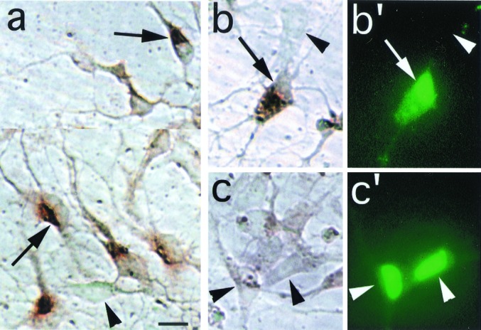
Ectopic expression of PAC1 receptor in cortical precursors. (a) The majority of normal, untransfected cortical precursors express PAC1 immunoreactivity (as previously reported in ref. 10) revealed by extended peroxidase incubation time (5 min, DAB substrate). Arrows, PAC1-IR cells exhibiting intense cytoplasmic signal and a negatively stained, eccentric nucleus; arrowheads, negative cell exhibiting only background signal. (b and b′) Following a brief (1 min) reaction time, this oblong cell cotransfected with GFP and PAC1 expression vectors exhibits intense cytoplasmic immunostaining for PAC1 (b, arrow) and prominent GFP autofluorescence in the eccentric nucleus (b′), although weaker fluorescence is present in the cytoplasm. (c and c′) In contrast, cells transfected with GFP vector alone exhibit intense GFP signal (c′), but only background DAB signal (c, arrowhead). Note that, in contrast to the GFP immunostaining procedure (Figs. 1 and 2), GFP autofluorescence is predominantly observed in the nucleus. (Scale bar = 10 μm.)