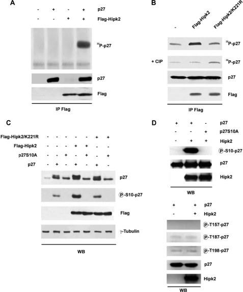
FIGURE 2.
HIPK2 phosphorylates p27kip1 at serine 10. A, FLAG-HIPK2 was overexpressed in HEK293 cells, TCEs were immunoprecipitated with anti-FLAG Ab, and kinase assays were performed on recombinant p27kip1 protein in the presence of [γ-32P]ATP. Kinase reaction products were resolved by SDS-PAGE and analyzed by autoradiography (upper panel). WB of immunocomplexes was performed with the indicated Abs (lower panels). B, TCEs from HEK293 cells transfected with FLAG-HIPK2 or its KD mutant, FLAG-HIPK2/K221R, were immunoprecipitated (IP) with anti-FLAG Ab. The immunocomplexes were incubated with recombinant p27kip1 protein in the presence of [γ-32P]ATP and then treated with calf intestinal phosphatase (CIP). In A and B, WBs with anti-FLAG Ab were performed on the immunoprecipitates as a control for protein loading. C, HEK293 cells transfected with the indicated plasmids and analyzed by WB for the expression levels of HIPK2, its KD mutant, p27kip1, and p27kip1 phosphorylated at Ser10. γ-Tubulin expression shows equal loading of samples. D, the effects of recombinant HIPK2 on phosphorylation of p27kip1 in vitro are shown. Upper panel, in vitro kinase assay with active recombinant HIPK2 incubated with p27kip1 or p27S10A recombinant proteins. The samples were analyzed by WB using the indicated Abs. Lower panel, in vitro kinase assays were performed incubating active recombinant HIPK2 with p27kip1 recombinant protein. Samples were analyzed by WB using specific Abs for the p27kip1 phosphorylated forms at threonine 157, threonine 187, and threonine 198. For A, B, C, and D, one representative of three independent experiments is shown.

