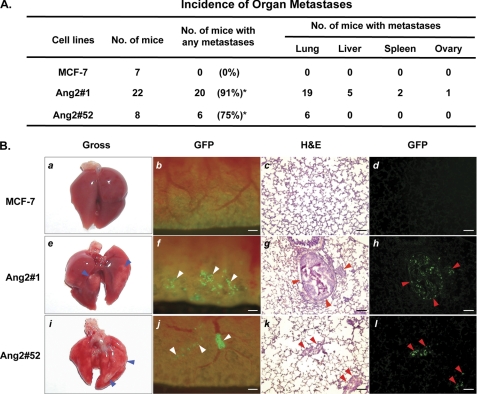
FIGURE 1.
Expression of exogenous Ang2 enables MCF-7 breast cancer cells to metastasize to several organs of mice. A, summary of the incidence of organ metastases in mice that separately received MCF-7 Ang2-expressing (Ang2#1 or Ang2#52) or GFP-expressing (MCF-7) cells. Fisher's exact test showed significantly higher incidence of metastasis in mice received Ang2#1 cells (*, p < 0.0001) or Ang2#52 (*, p = 0.007) as compared with mice received MCF-7/GFP cells. Data are combined from two independent experiments with 3–11 mice per group. B, representative images of the lungs of mice in A analyzed by different methods. MCF-7 breast cancer metastases in the lung of mice were examined 12–14 weeks after tail-vein injection by gross observation (panels a, e, and i) and epi-fluorescent observation of GFP (panels b, f, and j). Cryo-sections were further examined by epi-fluorescence (panels d, h, and l) followed by H&E staining (panels c, g, and k). Mice that received Ang2 #1 or Ang2#52 cells showed lung metastases (panels e to l) with a high incidence. Grossly visible metastases (blue arrowheads) in the lung (panels e and i) were identified as green foci (panels f and j, white arrowheads) and cells expressing GFP in the nuclei (panels h and l, red arrowheads), and further confirmed by H&E staining (panels g and k, red arrowheads). Scale bars: panels b to d, f to h, and j to l, 120 μm. Data are representative from two independent experiments shown in A with similar results.

