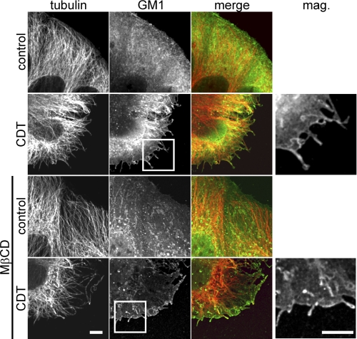
FIGURE 7.
Microtubule-based protrusions and the site of their formation are enriched in ganglioside GM1. Indirect immunofluorescence of α-tubulin (red) and GM1 staining by FITC-conjugated cholera toxin subunit B (green) in Caco-2 cells are shown. Cells were treated with 200 ng/ml CDTa and 400 ng/ml CDTb for 1.5 h or additionally pretreated with 5 mm MβCD to deplete cholesterol. Protrusions are labeled with cholera toxin. MβCD-treated cells form much less protrusions. The residual protrusions still have increased levels of GM1. Scale bars, 5 μm.

