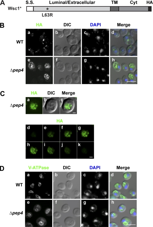
FIGURE 1.
Wsc1* localizes to the vacuolar lumen. A, schematic representation of Wsc1*. The L63R mutation is indicated by the asterisk, and the position of the HA epitope tag is shown in black. S.S., signal sequence; TM, transmembrane domain; Cyt, cytoplasmic domain. B, Wsc1* in wild type and Δpep4 cells was localized by indirect immunofluorescence using anti-HA monoclonal antibody and Alexa Fluor 488 goat anti-mouse antibody (green channel). Nuclear DNA was stained by DAPI to indicate positions of nuclei (blue channel). Visualization was performed using confocal and DIC microscopy as indicated. Scale bar, 5 μm. C, Wsc1* localization in Δpep4 cells as described in B. A z-series was captured, with the middle plane, its corresponding DIC image, and merged images shown in a–c. d–k show individual z-stacks of the series from top to bottom. Scale bars, 1 μm. D, wild type and Δpep4 cells were processed as in B and bound with anti-V-ATPase (60-kDa subunit) antibody as a vacuolar membrane marker followed by Alexa Fluor 488 goat anti-mouse antibody. Cells were visualized by confocal and DIC microscopy. Scale bar, 5 μm.

