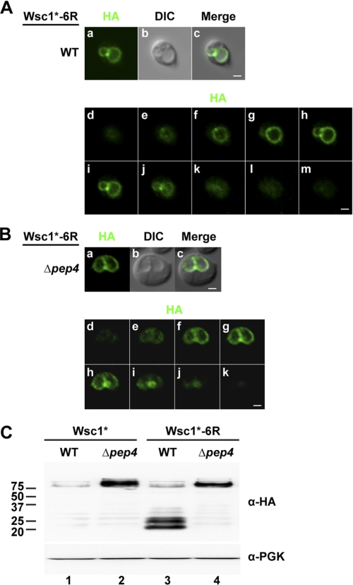
FIGURE 4.
A Wsc1* ubiquitination-deficient variant fails to enter the MVB pathway. Wsc1*-6R localization in wild type (A) and Δpep4 cells (B). Images in a–c are from the middle plane of a z-series displaying Wsc1* (HA), vacuoles (DIC), and their merged images. d–m (A) and d–k (B) show a series of z-stacks from top to bottom. Scale bars, 1 μm. C, Wsc1* and Wsc1*-6R expression in wild type and Δpep4 cells analyzed by immunoblotting using the anti-HA antibody.

