FIGURE 6.
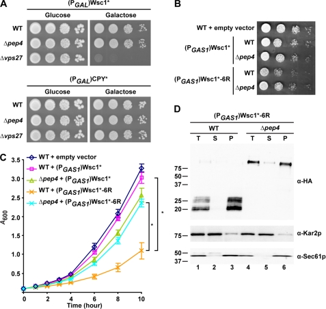
Accumulation of Wsc1* fragments causes toxicity. A, wild type, Δpep4, and Δvps27 cells containing (PGAL1)Wsc1* or (PGAL1)CPY* grown in raffinose media were spotted as 10-fold serial dilutions onto glucose (promoter-repressed) and galactose (promoter-activated) medium plates and incubated at 30 °C for 2 and 3 days, respectively. B, wild type and Δpep4 cells containing the control vector, (PGAS1)Wsc1*, or (PGAS1)Wsc1*-6R were spotted on selective synthetic plates and incubated at 30 °C for 2 days. C, growth analysis of wild type and Δpep4 cells containing the empty vector (pRS315), (PGAS1)Wsc1*, or (PGAS1)Wsc1*-6R. Cells were grown in selective synthetic media to log phase and diluted to 0.1 A/ml. Growth was measured as 0.1 A600/ml readings from cultures at regular intervals over 10 h. The data plotted reflect three independent experiments with the mean ± S.D. (error bars) indicated. *, p < 0.01, Student's t test. D, membranes prepared from wild type and Δpep4 cells expressing (PGAS1)Wsc1*-6R were treated with 0.1 m sodium carbonate, pH 11.0, for 30 min on ice. A portion was reserved as total (T), and the remaining was subjected to centrifugation at 100,000 × g. Supernatant (S) and membrane pellet (P) fractions were collected and analyzed by immunoblotting. Wsc1*-6R was detected using anti-HA antibody. Kar2p and Sec61p serve as soluble and integral membrane protein controls, respectively.

