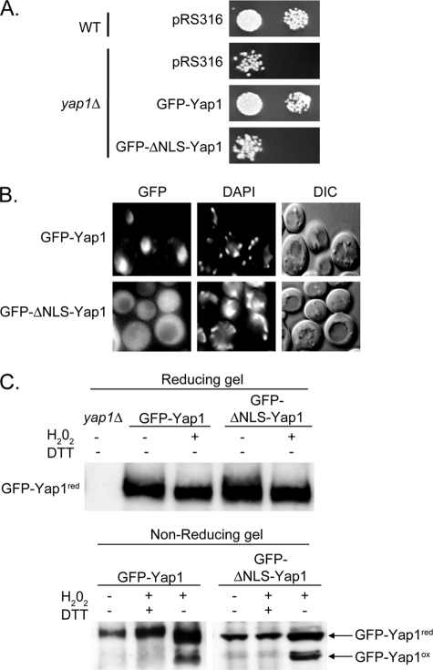
FIGURE 8.
Evidence for H2O2-induced folding of Yap1 occurring in the cytoplasm. A, wild-type or yap1Δ cells were transformed with a low-copy number vector plasmid (pRS316) or the same plasmid expressing GFP-Yap1 or the NLS-deficient mutant (GFP-ΔNLS-Yap1). Transformants were placed on rich medium containing H2O2 to compare their ability to tolerate H2O2 as before. B, the transformants containing the two indicated forms of GFP-Yap1 were treated with H2O2 for 10 min and then visualized by microscopy for localization of GFP-Yap1 (GFP), organellar DNA (DAPI), or by Nomarski optics (DIC). C, protein extracts were prepared under conditions allowing detection of the H2O2-folded form of GFP-Yap1 as described above. In the top panel, these extracts were resolved using standard SDS-PAGE (Reducing gel). In the bottom panel, a nonreducing gel was used to electrophorese proteins prepared under the indicated conditions. DTT was included in some samples to fully reduce GFP-Yap1. The locations of the reduced and oxidized forms of GFP-Yap1 are indicated as before. Both Western blots were probed with anti-Yap1 antibody.

