FIGURE 3.
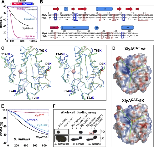
Designing a gain-of-function lysin (amidase) catalytic domain, XlyA+5KCAT. A, bactericidal activities of wild-type lysins and a chimera (XlyACAT:PlyLCBD) against B. cereus (Bcer) or B. subtilis (Bsub). B, sequence alignment and secondary structure assignments of XlyACAT and PlyLCAT. Residues involved in catalysis and substrate recognition are boxed in red. Residue pairs in blue boxes indicate sites mutated to lysine in XlyACAT to create XlyACAT+5K. Below the sequences, asterisks indicate identity; and colons indicate close similarity. C, stereo Cα overlay of XlyACAT (pink), Xly A+ 5KCAT (green), and PlyLCAT (cyan). XlyA mutated residues are labeled, and their side chains shown as sticks. Orientation is the same as in Fig. 2B. D, electrostatic surfaces of wild-type XlyACAT and XlyA+5KCAT, viewed as in A, showing that mutations do not affect the active site face. E, bactericidal activities (against B. subtilis) of wild-type and mutant XlyA catalytic domains, compared with full-length enzyme. XlyA+4KCAT is similar to XlyA+5KCAT but has one less residue changed to lysine. F, pulldown (PD) binding assays (Western blots using anti-His tag antibody) using whole cells (B. cereus, B. subtilis, or B. anthracis Sterne) and catalytically inactivated His-tagged mutants of PlyLCAT, XlyACAT, and XlyA5KCAT. LC = loading control. The input protein concentration was 1 μm or (for B. anthracis) 10 μm.

