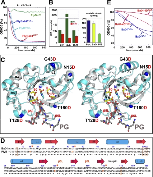
FIGURE 4.
Engineering a loss-of-function (muramidase/lysozyme) lysin. A, comparison of the lytic activities of full-length and catalytic domains of B. anthracis phage lysozymes, PlyBa04 and PlyB, against B. cereus ATCC 4243. The data for PlyB are derived from Fig. 1 of Porter et al. (47). Enzyme and bacterial concentrations and strains are identical. B, PlyBa04CAT and PlyLCAT have similar host ranges and synergize in killing B. cereus. Left panel, PlyBa04CAT is a highly effective killer of B. cereus, B. megaterium, and B. subtilis (t½ = 50–100 s with 0.8 μm lysin), similar to the behavior of PlyLCAT (and faster than the full-length protein in each case). Right panel, when used in combination, there is a clear synergistic effect between PlyLCAT and PlyBa04CAT against B. cereus. Total lysin concentration is the same in each case (0.4 μm). P/B = 0.2 μm PlyLCAT + 0.2 μm PlyBa04CAT). C, stereo overlay of PlyBa04 (cyan) and PlyB (gray) catalytic domains, with secondary structure elements as in C. The four mutation sites in PlyBa04CAT are indicated. The two pairs of conserved acidic residues at the active site are shown as ball-and-stick (PlyBa04 numbering), as is a molecule of buffer (MES), which lines the center of the site and may be a substrate mimetic. A glycan-muropeptide fragment is modeled based on an overlay with the structure of pneumococcal phage lysin, Cpl-1 (Protein Data Bank code 1H09), in complex with a fragment of PG. E, comparison of lytic activities of PlyBa04 wild-type and mutant lysins against B. cereus in the context of full-length and catalytic domains. D, structure-based sequence alignment and secondary structure assignments of PlyBa04CAT and PlyBCAT, with active residues boxed. Mutations sites are numbered and labeled with *.

