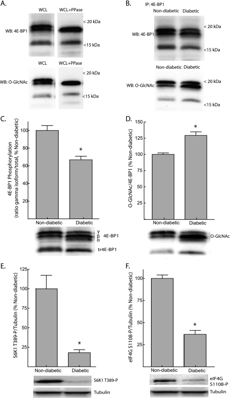
FIGURE 1.
Reduced phosphorylation and increased O-GlcNAcylation of 4E-BP1 in the liver of Ins2Akita/+ mice. A, Western blot analysis of supernatants from liver whole cell lysates (WCL) using anti-4E-BP1 (top panels) or anti-O-GlcNAc (bottom panels) antibodies, before (left panels) or after (right panels) treatment with λ-phosphatase (PPase). B, 4E-BP1 was immunoprecipitated (IP) from liver supernatants of non-diabetic or diabetic mice and subsequently subjected to Western blot (WB) analysis for 4E-BP1 (top panel) or O-GlcNAc (bottom panel) content. 4E-BP1 phosphorylation (C) and O-GlcNAcylation (D) in the liver of Ins2Akita/+ diabetic mice was assessed by Western blot analysis. Phosphorylation was assessed as the proportion of the protein present in the γ-form relative to the total amount of 4E-BP1 in all forms (α+β+γ). Activity of mTORC1 was assessed by Western blot analysis of S6K1 phosphorylation on Thr389 (E) and eIF4G on Ser1108 (F). Representative blots are show. Values are means + S.E. (n = 5). *, p < 0.05 versus non-diabetic.

