FIGURE 6.
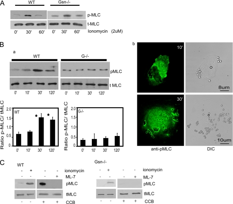
A, cells treated with ionomycin exhibit myosin light chain phosphorylation in WT and gelsolin null cells. B, a, bead-associated proteins collected from cells treated with collagen-coated beads show maximal phosphorylation of MLC at 30 min in WT cells but not in gelsolin-deficient cells. The histogram below shows ratios of quantification of blot densities as indicated. Data are mean ± S.E. of ratios. b, immunostaining shows localization of phospho-MLC (pMLC) at the bead sites after 10 and 30 min incubation with collagen-coated beads. C, in the presence of the MLC kinase inhibitor ML-7, gelsolin WT or null cells incubated with collagen beads show a complete block of MLC phosphorylation. The experiment was repeated three times. DIC, differential interference contrast; CCB, collagen-coated beads; tMLC, total myosin light chain.

